![An Australian Government Initiative [logo]](/images/austgovt_brown_90px.gif)

Reproduction & dispersal
Sexual Reproduction
Sexual reproduction involves the mixing of genes from two different parents to give offspring with a genetic make-up similar to, but different from, each parent. In bryophytes the process requires the production of male gametes (sperm), female gametes (eggs) and some means of getting the sperm to the eggs. The gametes are produced on the gametophytes. The sperm are produced within tiny, typically stalked, club-shaped structures called antheridia and you can also see bryophyte sperm referred to as antherozoids. The stalk anchors the antheridium to the gametophyte. Each antheridium produces numerous sperm. The eggs are produced in tiny, typically somewhat flask-like structures called archegonia. Each archegonium holds one egg (in a swollen section called the venter) and the sperm enter through the channel in the narrower, tubular section (or neck). On the side of the venter opposite the neck is the foot which anchors the archegonium to the gametophyte. In the early stages of archegonial development that channel does not exist, the area being filled with cells. At maturity the cells in the centre of the neck disintegrate to create the channel. The channel is filled with mucilage that results from the breaking down of the cells that initially occupied the channel.
A fertilized egg in an archegonium develops into the sporophyte. The sporophyte consists of a spore-containing capsule which, depending on the species, may be stalked or stalkless. Each spore contains a mix of genes from the two parents and on successful germination will give rise to a new gametophyte.
The following diagrams show some moss archegonia and antheridia. The figures have been copied from John Lindley's The Vegetable Kingdom, published in 1853. The archegonia are on the left and have been coloured green You can see the swollen venters near the archegonial bases. At the top of the neck each archegonium has a somewhat funnel-shaped mouth. The antheridia are on the right, have been coloured blue and the middle antheridium is releasing a sperm mass, coloured brownish-orange. Archegonia and antheridia grow intermixed with hair-like to club-like paraphyses, left uncoloured in the diagrams. The previous paragraph mentioned that the antheridia and archegonia are tiny. Size varies, depending on species, but typically these gamete-producing organs are well under a millimetre in length.
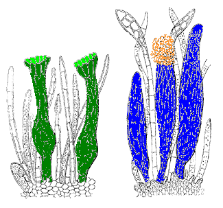
Bryophyte antheridia are fairly uniform in structure and the same is true for the archegonia. The antheridia vary in size and shape (from globose to somewhat cylindrical) depending on species, but the diagram above captures the essence of any antheridium - a short, narrow stalk supporting a swollen, sperm-producing organ. Similarly, the archegonia vary in size, and relative lengths of the neck, the venter and the length of the supporting foot - but the diagram above shows the essential features of all archegonia.
Individual antheridia and archegonia are microscopic but at times you can see where they are formed. In this photo ![]() of the moss Rosulabryum billardieri each yellow ball is a cluster of antheridia. The same is the case in this photo
of the moss Rosulabryum billardieri each yellow ball is a cluster of antheridia. The same is the case in this photo ![]() of a thallose liverwort in the genus Fossombronia. This photo
of a thallose liverwort in the genus Fossombronia. This photo ![]() shows male plants of the hornwort Phaeoceros inflatus and antheridia are produced in the many "blisters" visible on the thallus. Groups of archegonia are found under the white "blisters" shown in this photo
shows male plants of the hornwort Phaeoceros inflatus and antheridia are produced in the many "blisters" visible on the thallus. Groups of archegonia are found under the white "blisters" shown in this photo ![]() of the thallose liverwort Lunularia cruciata.
of the thallose liverwort Lunularia cruciata.
Though there is much uniformity at the structural level, there is variety in the formation and arrangement of the archegonia and antheridia. The rest of this page will give an overview of the sexual reproduction cycle![]() .
.
Getting sperm to egg
Once an antheridium has matured and contains viable sperm, the sperm need to get to the eggs in archegonia. The first step for the sperm to get out of the antheridia and the second is to then travel to the archegonia and fertilize the eggs within. Water is essential for both steps.
In some bryophytes a mature antheridium will hold free sperm, but more commonly that's not the case, Rather, each sperm is still held within the cell in which it formed. In such a case, when an antheridium opens, those sperm-containing cells are released and it is only at some stage after release from the antheridium that the single sperm, within each such cell, is liberated. Such liberation may take place shortly after the opening of the antheridium or as long as 15 minutes later, depending on species. In the following, the expression "sperm mass" will mean either a mass of free sperm or a mass of sperm-containing cells, when it's not essential to distinguish the two.
When a mature antheridium is moistened the cells at the apex absorb water, swell and finally burst or open in some way. The sperm mass inside a mature antheridium is under pressure. So, once an antheridium has opened, the sperm mass is forced out. In some bryophytes the force is enough to shoot the sperm mass into the air, allowing dispersal over a relatively wide area. However, in most cases the sperm mass simply oozes into the area around the antheridium and further dispersal is by some other means. While the entire sperm mass may sometimes be released during the forceful extrusion, release is more often a two-stage process. Typically a large percentage of the spore mass is quickly forced out by the built-up internal pressure, but a proportion remains within the antheridium and exits more slowly, over many minutes.
The summary in the previous paragraph is enough to give you a quick grasp of the broad features of the process, but there is variation in the finer detail between species. The LIBERATION & DISPERSAL OF SPERM page looks more closely at some of the steps in a few bryophytes. The sperm-to-egg process has been thoroughly studied in a relatively small number of bryophytes. Thus, the examples given on that page may not explain the processes in all bryophytes, but you will at least see some of the variations that are known to occur![]() .
.
After fertilization
Once an egg has been fertilized the development of the sporophyte begins. The fertilized egg elongates and after a few cell divisions begins to differentiate. The lower portion usually becomes a foot that penetrates the gametophyte and anchors the embryonic sporophyte to the gametophyte. The upper will develop into the spore-bearing capsule (and also the supporting stalk or seta, in species in which the mature capsule is stalked).
The sporophytes are at least partially dependent on the gametophyte for nutrients. Transfer cells develop at the sporophyte-gametophyte boundary in the majority of bryophytes, but not all. These cells are specialized cells that allow efficient transfer of nutrients from the gametophyte to the sporophyte. In the bryophytes where they do occur they may be formed on the gametophyte, the sporophyte or both. So, combined with the possibility of no transfer cells, there are four possibilities. All hornworts have transfer cells and they form only on the gametophyte. Three of the four possibilities occur in mosses. In the majority of moss genera the transfer cells are found on both the gametophyte and the sporophyte, though they are absent in Sphagnum and in a small number of moss genera they are found only on the sporophyte. A common example of the last is the genus Polytrichum and its close relatives. In the liverworts all four possibilities occur. The leafy liverworts have transfer cells only on the sporophytes. In the complex thallose liverworts the transfer cells are found on both sporophyte and gametophyte. In the simple thalloid liverworts there are examples of all four possibilities.
The gametophyte-sporophyte junction often has a convoluted, maze-like form. This gives a larger surface area (and hence more transfer cells) than would a simple, smooth boundary and so increases the rate at which nutrients can flow to the sporophyte![]() .
.
After fertilization, the archegonium becomes modified into a protective sheath around the young sporophyte. There are significant differences, in both structure and development, between hornwort, liverwort and moss sporophytes. You can find out more about the external appearance by going to the BRYOPHYTE GROUPS SECTION. In the SPOROPHYTE DEVELOPMENT SECTION you'll find more detailed accounts of sporophyte development and their internal structure.
In mosses the archegonia are typically formed in groups. In many cases once one archegonium in such a group has been fertilized the others lose the ability to be fertilized. This appears to be caused by an inhibitory hormone released from a fertilized archegonium. In such circumstances only one sporophyte can develop from that archegonial group. However, in some circumstances more than one sporophyte may develop from an archegonial group. Such a phenomenon is called polysety. It may be due to two archegonia being fertilized simultaneously or perhaps because of too low an amount of inhibitory hormone being produced.
See also:
Liberation & dispersal of sperm
Vegetative Reproduction
Sexual vs Vegetative Reproduction