![An Australian Government Initiative [logo]](/images/austgovt_brown_90px.gif)

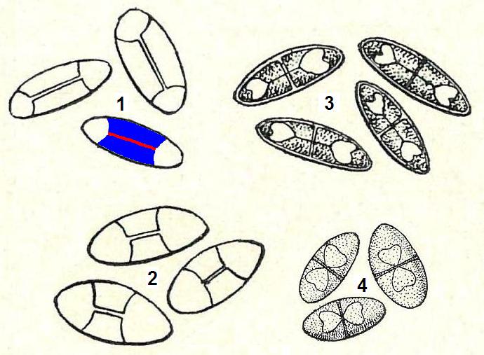
Spores - some more septate forms (from Albert Schneider's A text-book of general lichenology)
The spores in illustrations 1 and 2 are examples of polarilocular spores. There are two cells (or locules) at opposite ends of the spore, separated by a thick septum but one in which a connecting channel is easily visible in a light microscope. The opposite ends of an elongated spore are sometimes referred to as the poles of the spore and this explains the origin of the term polarilocular, since the locules are at the poles. I have added colour to one of the drawings to make the polarilocular spore structure easier to understand. Red indicates the connecting channel between the two (white) polar locules and the thick septum is coloured blue. The two other spores have multi-angular locules, two per spore.
1. Theloschistes chrysophthalmus (note that today's spelling of the genus name has no h after the initial t); 2. Placodium elegans; 3. Pyxine picta; 4. Rinodina sophodes.
Note: not all to the same scale.
Albert Schneider's A text-book of general lichenology, published in Binghamton in 1897 by Willard N. Clute & Co. illustrates the structural features of various lichens from the north-eastern United States.