![An Australian Government Initiative [logo]](/images/austgovt_brown_90px.gif)

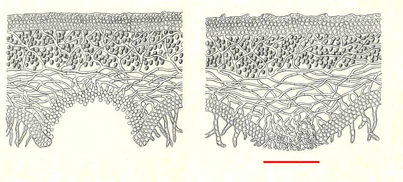
Cyphella and pseudocyphella (from Albert Schneider's A text-book of general lichenology)
Both illustrations show cross sections through thalli. In each cross section you can see a well-developed upper cortex, then the photobiont layer and the loose weave of the medullary hyphae and finally a well-defined lower cortex with short rhizines extending from the lower cortex.
In the left hand illustration you can see a cyphella, its recessed surface formed by a "pseudo cortex" of cells markedly different from both those of the lower cortex and the hyphae of the medulla. On the right (indicated by the red bar) is a slightly convex pseudocyphella, where medullary hyphae protrude through a gap in the lower cortex. Those hyphae, though well-branched, are essentially undifferentiated from the rest of the hyphae in the medulla
Albert Schneider's A text-book of general lichenology, published in Binghamton in 1897 by Willard N. Clute & Co. illustrates the structural features of various lichens from the north-eastern United States.