![An Australian Government Initiative [logo]](/images/austgovt_brown_90px.gif)

") Episodes in Australian lichenology - After the first century
Episodes in Australian lichenology - After the first century
Rex Filson and Mac.Robertson Land
Rex Filson's monograph The Lichens and Mosses of Mac.Robertson Land was published in 1966 by the Antarctic Division of the Australian Department of External Affairs. Here is an extract from the abstract at the beginning of the book:
The distribution of lichens and mosses in Mac.Robertson Land is recorded and discussed, and workable descriptions of all the known species in this region have been prepared...A complete list of the present and previous collections in Mac.Robertson Land has been compiled and some comments have been offered.
This work was by no means the first published study of Antarctic lichens but it was the first scientific monograph on the subject to be published with colour illustrations, namely a number of Filson's excellent paintings. His colour paintings of lichens show the features you'd see with a naked eye or a hand lens. Here, taken from the book's Plate 5, is a detail showing a few black apothecia of Buellia frigida on white thalli. The brown area is the underlying rock and the scale bar represents one millimetre.
 plate 5
A taller lichen is the fruticose species Usnea antarctica, shown here growing on a rock, with the scale bar representing one centimetre.
 plate 36
Apart from the colour illustrations the book contained many monochrome illustrations. Sometimes these showed external features such as the following which shows the structure of an anchoring, superficially root-like rhizine from a specimen Filson had identified as Omphalodiscus antarcticus. If you'd like to see this species in colour, look here. PLATE 33
Filson also showed microscopic detail. For example, in the following series of four figures, the two on the left illustrate some features of Lecanora expectans. The first shows a forked paraphysis and to its right a depiction of part of an apothecium in which you can see several asci in different stages of development. Paraphyses (the plural of paraphysis) are sterile hyphal ends that are present in the apothecium amongst the asci. They vary greatly in shape and colour between species, sometimes being no more than un-modified hyphae while at other times they may be forked or have swollen ends. They may be uncoloured or pigmented, smooth or encrusted, septate or with no internal divisions.
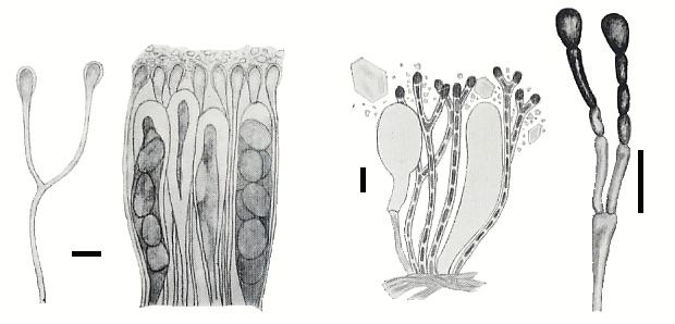
The third figure shows several paraphyses ( as well as two asci and a scattering of granules) of Biatorella antarctica and rightmost is a branched paraphysis of Buellia grimmiae. Each scale bar represents 10 micrometres. The Buellia paraphyses are septate slightly constricted at the septa. The book featured the first description of Buellia grimmiae and here PLATE 9 is the plate showing the microscopic features of the species.
History of Australian lichenology pages on this website |