![An Australian Government Initiative [logo]](/images/austgovt_brown_90px.gif)

 Fungal
ecology
Fungal
ecology
Fungi and invertebrates
There are numerous fungus-invertebrate associations, many of which are discussed
in the reference given in the button and this link [http://www.botany.hawaii.edu/faculty/wong/BOT135/lect24.htm]
![]() discusses
a few examples of fungus-insect symbioses.
discusses
a few examples of fungus-insect symbioses.
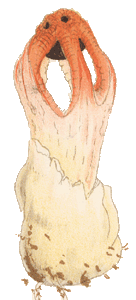
The aptly named Stinkhorn fungi rely on carrion-loving insects for spore-dispersal.
One thing common to all Stinkhorns is the production of spores in a foul-smelling,
khaki to brownish slime. You can see this spore-bearing slime at the top of
this Dictyophora multicolor ![]() .
The smell is a powerful attractant of various flies and midges. In some other
Stinkhorns, such as Anthurus archeri
.
The smell is a powerful attractant of various flies and midges. In some other
Stinkhorns, such as Anthurus archeri ![]() and Aseroë rubra
and Aseroë rubra ![]() ,
there is an additional colour cue. The red in these Stinkhorns looks much like
freshly exposed meat.
,
there is an additional colour cue. The red in these Stinkhorns looks much like
freshly exposed meat.
In tropical America stingless bees in the genus Trigona have been seen
collecting the exudates (and possibly spores) from stinkhorns in the genera
Dictyophora and Phallus. Entomologists studying these examples
found no exudate or spores on the bodies of the bees, strongly suggesting that
the bees swallowed the exudate and spores. ![]() Presumably
the bees include spores in their diet. It is also possible that at least some
spores will pass, undigested, through the bees - in which case the bees would
act as dispersal agents for the fungi.
Presumably
the bees include spores in their diet. It is also possible that at least some
spores will pass, undigested, through the bees - in which case the bees would
act as dispersal agents for the fungi.
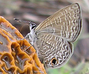 The butterfly Prosotas dubiosa on fungus Dictyophora multicolor |
In late March 2006 the Australian National Botanic Gardens received an enquiry about a fungus found growing in a garden at Moore Park Beach, 25 km north of Bundaberg, Queensland. The fungus was Dictyophora multicolor and that very specimen is shown here ![]() . What was particularly interesting was that the photo showed a butterfly on the upper-right of the Stinkhorn. From a closer look, at an enlargement of the photo, it was clear that the butterfly's proboscis, though a little fuzzy, was uncoiled and with its further end either in contact with, or very close to, the fungal surface. The angle makes it hard to tell. There's an enlargement of the butterfly here (right). This was an interesting photo because there appeared to be no other reports of any butterfly visiting a Stinkhorn. The photo was taken by Sandra Sullivan, who has very kindly allowed its reproduction on this website. She also noted that she had noticed a couple of small butterflies on the fungus. This suggests that the visitation could be more than just a chance event.
. What was particularly interesting was that the photo showed a butterfly on the upper-right of the Stinkhorn. From a closer look, at an enlargement of the photo, it was clear that the butterfly's proboscis, though a little fuzzy, was uncoiled and with its further end either in contact with, or very close to, the fungal surface. The angle makes it hard to tell. There's an enlargement of the butterfly here (right). This was an interesting photo because there appeared to be no other reports of any butterfly visiting a Stinkhorn. The photo was taken by Sandra Sullivan, who has very kindly allowed its reproduction on this website. She also noted that she had noticed a couple of small butterflies on the fungus. This suggests that the visitation could be more than just a chance event.
For a definitive identification of the butterfly a specimen or at least a view of the upper wing surfaces would be necessary. The photograph shows only the underside, since the wings are folded. On the basis of this limited evidence, CSIRO Entomology in Canberra noted that the closest match is with the genus Prosotas, of the family Lycaenidae. The wing details most closely match the species Prosotas dubiosa. This butterfly is known from Sri Lanka and India, through to northern and eastern Australia and the Solomon Islands. It is a species of humid, tropical or sub-tropical areas, but has also been found once at Yunta, in arid South Australia, in 1999![]() .
.
The South Australian record of Prosotas dubiosa is noted at: http://users.chariot.net.au/~rbg/dubiosa_ds.htm
The adult or juvenile stages of various flies, mites, gnats, beetles, springtails and nematodes eat fungal fruiting bodies, spores or mycelia. To give just one specific example, flies of the genus Tapeigaster are commonly seen on a variety of Australian mushrooms and boletes. Typically, a highly territorial male will patrol the cap of a mushroom or bolete while the female lays her eggs on the gills or pores below. Australian Tapeigaster species have been reared on a number of mushroom and bolete species.
If you take a medium to large mushroom cap and shake it gently (gills facing down) over a sheet of paper you are likely to end up with a collection of varied invertebrates on the paper. Amongst them will be a mixture of juvenile and adult forms with various feeding strategies. Some will be generalist feeders on any fungal tissue while others will be specialists, perhaps just feeding on the spores, for example. Then there will be those feeding on other invertebrates. The corky to woody bracket fungi are also often good harbours of invertebrates.
Various invertebrates have evolved associations with specific fungi. For example, in Central America the leafcutter ants farm fungi on digested leaf fragments. A very informative site is: http://www.zi.ku.dk/personal/drnash/atta/Pages/Leafcut.html
While many termites from Africa through to Asia also grow fungi but use the termite faeces as a base. The guts of these termites lack the wood-digesting microbes found in other termites and so the African-Asian species rely on the fungi for food.
The wood wasp Sirex noctilio was accidentally introduced to Australia
in the 1950s and has caused considerable economic loss in Australian pine plantations.
The larvae develop in trees that have been infected with a fungus in the genus
Amylostereum. The fungus may regulate the water content in wood, to a
level optimum for egg or larval development, or the larvae may eat the fungus.
When an adult female emerges from her pupal case she picks up fungal cells in
a pouch located near the base of her ovipositor. When she lays her eggs in another
tree she simultaneously squeezes out fungal cells from the pouch, thereby initiating
a new fungal infection. The fungus is carried to new host trees and seems to
do so well out of this arrangement that it does not produce fruiting bodies
in Australia. It has not lost the ability to do so, for fruiting bodies can
be artificially induced in the laboratory.![]() However, in the wild the fungus saves a considerable expenditure of resources
by relying on the wasp for dispersal. There are also a number of beetles which
have similar associations with fungi.
However, in the wild the fungus saves a considerable expenditure of resources
by relying on the wasp for dispersal. There are also a number of beetles which
have similar associations with fungi.
Vegetable caterpillars
Numerous fungi are parasitic on invertebrates, though many of them are microfungi
and so outside the scope of this website. However, Cordyceps is a widespread
macrofungal genus, with the bulk of the species being parasitic on invertebrates.
The adults or larvae of numerous moths, beetles, ants, wasps or spiders are
the victims, with the fungi killing their hosts. This link ![]() shows exquisite paintings of various species of Cordyceps and a couple
of other genera.
shows exquisite paintings of various species of Cordyceps and a couple
of other genera.
Cordyceps gunnii ![]() ,
,
![]() is a fairly common Australian species. One photo shows the above-ground fruiting
body of the fungus and the other shows the excavated larva and fungal fruiting
body. The mycelium is ordinarily out of sight, in a below-ground larva of Ghost
Moths in the genus Oxycanus. In this photo
is a fairly common Australian species. One photo shows the above-ground fruiting
body of the fungus and the other shows the excavated larva and fungal fruiting
body. The mycelium is ordinarily out of sight, in a below-ground larva of Ghost
Moths in the genus Oxycanus. In this photo ![]() there are some examples of another Cordyceps species. By the time the
club-like fruiting body is produced, the invertebrate has been dead for some
time, its body filled with mycelium, hence the common name of Vegetable Caterpillar
given to the genus Cordyceps.
there are some examples of another Cordyceps species. By the time the
club-like fruiting body is produced, the invertebrate has been dead for some
time, its body filled with mycelium, hence the common name of Vegetable Caterpillar
given to the genus Cordyceps.
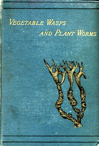 Other
names given to members of the genus are vegetable wasps and plant worms, also
the title of a book by the 19th century English mycologist MC Cooke.
The cover of the book, shown here, bears a drawing of Cordyceps taylori.
This is an Australian species, discovered in 1837 on the banks of the Murrumbidgee
River, 10 miles from Yass in NSW. In his book, Cooke called this the Murrumbidgee
Vegetable Caterpillar. It parasitizes the larvae of moths in the genus Trictena.
The smooth, antler-like areas are the above-ground fruiting bodies, while the
knobbly sections are the below-ground, parasitized larvae. Here
Other
names given to members of the genus are vegetable wasps and plant worms, also
the title of a book by the 19th century English mycologist MC Cooke.
The cover of the book, shown here, bears a drawing of Cordyceps taylori.
This is an Australian species, discovered in 1837 on the banks of the Murrumbidgee
River, 10 miles from Yass in NSW. In his book, Cooke called this the Murrumbidgee
Vegetable Caterpillar. It parasitizes the larvae of moths in the genus Trictena.
The smooth, antler-like areas are the above-ground fruiting bodies, while the
knobbly sections are the below-ground, parasitized larvae. Here ![]() is an extract from Cooke's book in which he discusses four Cordyceps species found in Australia. Three of the four had been discovered first in Australia whereas the first record of Cordyceps robertsii was from New Zealand.
is an extract from Cooke's book in which he discusses four Cordyceps species found in Australia. Three of the four had been discovered first in Australia whereas the first record of Cordyceps robertsii was from New Zealand.
Theodor Holmskjold's Beata ruris otia Fungis Danici has been said to contain the finest fungal illustrations published in pre-modern times. The illustrations were printed as engraved outlines and were then hand-coloured and one of the species illustrated was named Clavaria militaris by Holmskjold. He was the first to observe that the fruiting bodies grew on dead insects. The general belief had been that the dead insect somehow was transformed into the fungus. The species is now known as Cordyceps militaris and here ![]() is a link to a PDF of the Beata ruris plate for this fungus.
is a link to a PDF of the Beata ruris plate for this fungus.
Fungus sucks sap-suckers
The species in the genus Septobasidium form relationships with scale
insects. These photos show a couple of examples of the flattish Septobasidium
fruiting bodies ![]() ,
,
![]() ,
collected from plants in the Australian National Botanic Gardens. Septobasidium
is a worldwide genus, with about 200 species, and is fairly common in Australia.
,
collected from plants in the Australian National Botanic Gardens. Septobasidium
is a worldwide genus, with about 200 species, and is fairly common in Australia.
The photos show nothing unusual. However, if you look beneath that brown surface
you will find a number of scale insects. These insects are plant parasites and
suck plant sap through long tubes that the insect inserts into the plant. In
turn, Septobasidium grows over the insects, fungal hyphae penetrate the
insects and absorb nutrients from them. The scale insects are immobile and it
would seem that this is simply a case of parasitism with the fungus getting
all the benefits, but there are benefits to the embedded scale insects. ![]() They
are sheltered from the environment and protected from predators, allowing them
to live longer than their free-living brethren. It has been shown that, in at
least some cases, the fungal covering over the scale insects is deeper than
the reach of the ovipositors of parasitic wasps that seek out scale insects.
They
are sheltered from the environment and protected from predators, allowing them
to live longer than their free-living brethren. It has been shown that, in at
least some cases, the fungal covering over the scale insects is deeper than
the reach of the ovipositors of parasitic wasps that seek out scale insects.
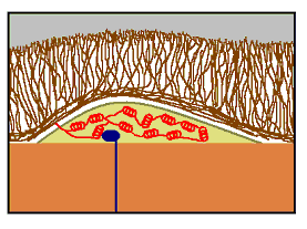 The
accompanying stylized diagram shows the basic features in a scale-Septobasidium
association. The scale insect is the centrally positioned khaki coloured object
and it’s sitting on the outside of a branch (coloured light brown). The
scale’s sucking tube (blue) penetrates deep into the plant. You can see
the numerous dark brown hyphae that make up the bulk of the Septobasidium.
Inside the scale are numerous coiled hyphae (in red), which are the haustoria
through which the fungus absorbs nutrients from the scale. In this case, the
fungus has entered the scale from just one point, to the left.
The
accompanying stylized diagram shows the basic features in a scale-Septobasidium
association. The scale insect is the centrally positioned khaki coloured object
and it’s sitting on the outside of a branch (coloured light brown). The
scale’s sucking tube (blue) penetrates deep into the plant. You can see
the numerous dark brown hyphae that make up the bulk of the Septobasidium.
Inside the scale are numerous coiled hyphae (in red), which are the haustoria
through which the fungus absorbs nutrients from the scale. In this case, the
fungus has entered the scale from just one point, to the left.
LINKS TO OTHER SECTIONS
-
Wood rotting fungi
-
Dung fungi
-
Fungi and invertebrates
-
Fungi and vertebrates
-
Fungi and fire
-
Fungi versus fungi
-
Mycorrhizas